The Use of the Buccal Fat Pad as a Pedicle Graft
This case highlights the use of the buccal fat pad as a pedicle graft for maxillary sinus closure.
-
Length: 14 Minutes
-
1 Video
About this video
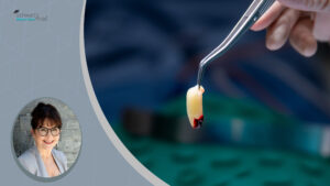
In this case, two failed dental implants had migrated into the maxillary sinus, resting within its cavity. Access to the sinus was achieved via a lateral wall approach, allowing for the removal of the displaced implants. To close the sinus and repair the perforated area, the buccal fat pad was mobilized by releasing it through the buccinator muscle and utilized as a pedicle graft. This technique provides structural volume, enhances vascularization, and ensures effective protection of the surgical site.
Related Classes
-
Length: 25 Minutes
-
1 Video
-
Length: 15 Minutes
-
1 Video
- Ridge preservation at the time of implant extraction.
- 3D ridge augmentation (vertical and horizontal) using an autologous onlay bone block graft.
-
Length: 42 Minutes
-
1 Video
Bilateral Inferior Alveolar Nerve Lateralization and Transposition
Lateralization and transposition of the mandibular canal with simultaneous implant placement for fixed prosthetic rehabilitation, including immediate implant placement and provisionalization.
Ridge Preservation, Sinus Augmentation, and Pedicled Buccal Fat Graft in an Infected Site Before Implantation in the Atrophic Posterior Maxilla
Bone augmentation, including ridge preservation and maxillary sinus augmentation, is performed in the posterior atrophic maxilla following the removal of an infected tooth.
Ridge Preservation and 3D Augmentation Following Failed Implant Removal
A two-step augmentation approach after failed implant removal: