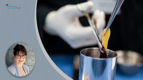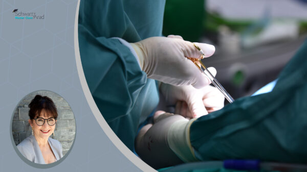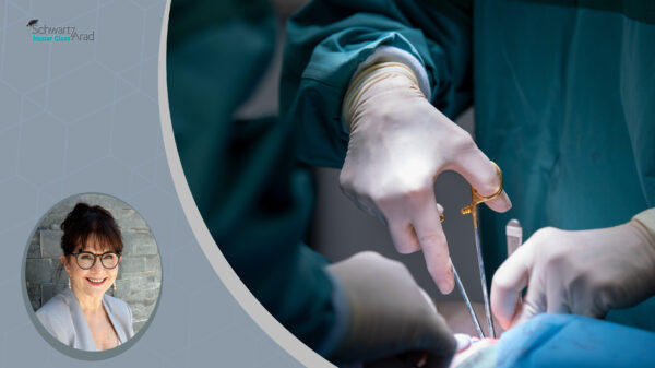
Mastering Severe Posterior Maxillary 3D Atrophy: Large Schneiderian Membrane Perforation Management with TAC and Multi-Modal onlay block bone grafts Augmentation
A demanding posterior maxillary reconstruction case demonstrating TAC-assisted repair of a large Schneiderian membrane perforation, combined with subnasal and sinus elevation procedures using autologous bone, bovine xenograft, and PRP to achieve significant horizontal and vertical bone augmentation in a severely atrophic posterior maxilla.
-
 Length: 42:41 Minutes
-
 1 Video
 Add to cart

About this video
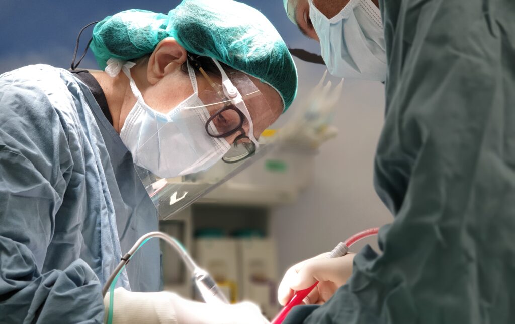
This narrated surgical video presents a comprehensive approach to one of the most challenging scenarios in implant-driven bone reconstruction: severe posterior maxillary atrophy complicated by a large intraoperative Schneiderian membrane perforation. The case demonstrates how TAC (tissue adhesive/collagen) is employed to achieve reliable membrane repair as a prerequisite for proceeding with augmentation in the same surgical session. The reconstruction protocol combines subnasal elevation with lateral window sinus augmentation, utilizing a blended grafting strategy of autologous bone and bovine-derived xenograft material enriched with PRP, targeting simultaneous horizontal and vertical bone volume gains across a partially edentulous posterior segment, covered with buccal pad of fat and PPP. The video addresses decision-making at each critical juncture — from perforation recognition and repair to graft layering rationale and flap management — providing clinicians with a replicable framework for managing complex augmentation cases where anatomical limitations and intraoperative complications converge. Intended for oral surgeons, periodontists, and implantologists engaged in advanced pre-implant surgical reconstruction.